| Video Discription |
Helicobacter pylori, is a gram-negative spiral-shaped bacterium, and it affects up to 50% of the population worldwide.
Up to 90% of people infected with Helicobacter pylori never experience symptoms or complications.
But in other part of population it causes several diseases: Peptic Ulcers; chronic or atrophic gastritis. And even cancers;
Individuals with chronic Helicobacter pylori infection have an increased risk of acquiring a cancer that is directly related to this infection. These cancers are stomach adenocarcinoma, and, less commonly diffuse large B-cell lymphoma of the stomach or other parts of the body.
Individuals infected with Helicobacter pylori, may also develop colorectal or gastric polyps. They are benign, but over time, especially for colorectal polyps, it can turn to colorectal cancer.
symptoms of Helicobacter pylori infection can be acute or chronic.
Acute symptoms include:
Abdominal Pain, Stomach ache, or nausea.
Chronic symptoms include:
Stomach pain, bloating, belching, nausea, sometimes vomiting. Such symptoms are called non-ulcer dyspepsia.
If ulcer develops it can have pain, which typically occurs when the stomach is empty, between meals, and in the early morning hours, but it can also occur at other times.
Even ulcer can be asymptomatic. Less common ulcer symptoms include nausea, vomiting, and loss of appetite.
Helicobacter pylori infection is usually acquired in early childhood and persists in the absence of treatment.
Helicobacter pylori harms stomach and duodenal linings by several mechanisms.
First, Ammonia produced by Helicobacter pylori, is toxic for cells. Also proteases which further damages cells. And cytotoxin, associated gene CagA can cause inflammation and can be carcinogen also.
Transmission:
Most common way of transmission of Helicobacter pylori is, oral-oral route. For example among family members who share utensils and kitchen tools during feeding.
Other common transmission is fecal – oral and sexual routes.
Colonization:
H. pylory colonization consist of 4 important steps:
1. urease activity of Helicobacter pylori, countering the acidic environment of the stomach. 2. Flaggela mediated motility, helps Helicobacter pylori move towards host gastric cells. 3. Adhesion to host cell receptors and 4. Successful colonization and persistent infection.
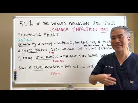
Diagnosis:
Helicobacter pylori infection is diagnosed by both invasive and non-invasive methods. Noninvasive tests include the detection of Helicobacter pylori antigens in the stool, detection of antibodies against Helicobacter pylori in serum, urine and oral samples, and a urea breath test (UBT).
The stool antigen test and Urea Breath Test have high sensitivity and specificity, similar to the invasive methods.
Invasive tests require gastric tissue for detecting the organism and include culture.
Culture is the only method with 100% specificity.
Urea breath test means, patient drinks 14C – or 13C-labelled urea, which the bacterium metabolizes, producing labelled carbon dioxide that can be detected in the breath).
By Yutaka Tsutsumi, M.D.ProfessorDepartment of PathologyFujita Health University School of Medicine - Yutaka Tsutsumi, M.D.ProfessorDepartment of PathologyFujita Health University School of Medicinehttp://info.fujita-hu.ac.jp/~tsutsumi/photo/photo002-6.htmhttp://info.fujita-hu.ac.jp/~tsutsumi/image/002/2-6.jpg, Copyrighted free use, https://commons.wikimedia.org/w/index.php?curid=535442
By User:KGH - Own work, CC BY-SA 3.0, https://commons.wikimedia.org/w/index.php?curid=507399
By BruceBlaus - Own work, CC BY-SA 4.0, https://commons.wikimedia.org/w/index.php?curid=44923645
By Amadalvarez - Own work, CC BY-SA 4.0, https://commons.wikimedia.org/w/index.php?curid=47205207
By Ed Uthman from Houston, TX, USA - Spleen, Diffuse Large B Cell LymphomaUploaded by CFCF, CC BY 2.0, https://commons.wikimedia.org/w/index.php?curid=30104814
By Polyp.jpeg: Original uploader was Rsabbatini at en.wikipediaLater version(s) were uploaded by Kd4ttc and dr. F.C. Turner at en.wikipedia.derivative work: Dr. F.C. Turner (talk) - Polyp.jpeg, CC BY 2.5, https://commons.wikimedia.org/w/index.php?curid=7057301
By Dowland58 - endoscopic picturePreviously published: never, CC BY-SA 3.0, https://commons.wikimedia.org/w/index.php?curid=22088176
By Polyp.jpeg: Original uploader was Rsabbatini at en.wikipediaLater version(s) were uploaded by Kd4ttc and dr. F.C. Turner at en.wikipedia.derivative work: Dr. F.C. Turner (talk) - Polyp.jpeg, CC BY 2.5, https://commons.wikimedia.org/w/index.php?curid=7057301
[H378ukXBS44] |